






3D Volume Imaging Wireless Ultrasound: Features That Matter
- 2026-11-10
- 53
- Guangzhou Sonostar Technologies Co., Limited
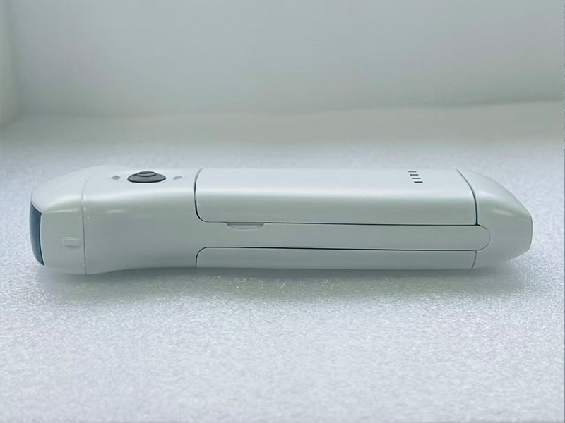
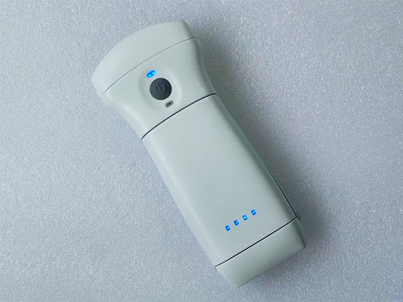
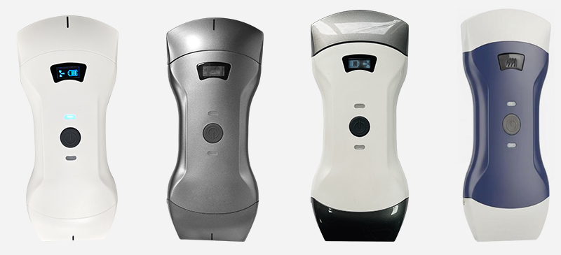
3D volume imaging wireless ultrasound combines two things that used to sit in separate price brackets: volume reconstruction, which lived on cart-based systems, and a probe that weighs 325 grams and talks to a phone over Wi-Fi. What determines whether such a device is genuinely useful is not the word “3D” on the box but a short list of specifications — sector geometry, depth settings, the volume toolbox, power architecture and export format.
Applications the features actually unlock
Obstetric triage and counselling. A 60° sector with depths to 305 mm is enough to render a fetal profile or a limb at the point of contact, and to show the patient what the clinician is describing. It supports triage and communication; it does not replace an anomaly scan on a high-end system, and positioning it as such invites trouble.
Abdominal organ assessment. Kidney, bladder and focal liver lesions become easier to discuss once the operator can cut a plane through them. The multiple depth settings mean the same probe covers a superficial gallbladder and a deep kidney in one session.
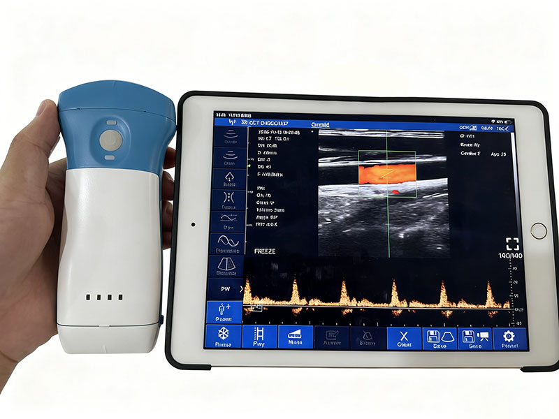
Musculoskeletal and pain intervention. On the line-array version, 2–20 MHz reaches tendon, fascia and small joints, and a volume showing lesion depth relative to the planned needle path shortens the planning conversation.
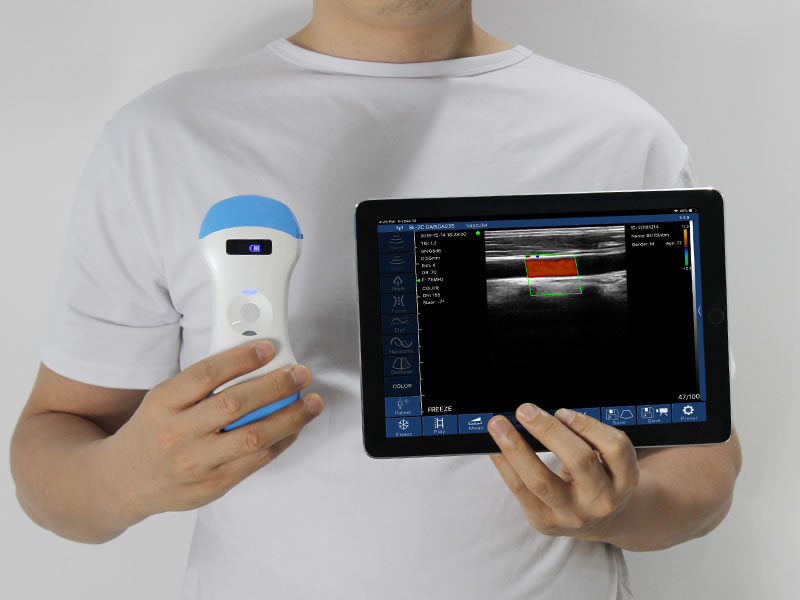
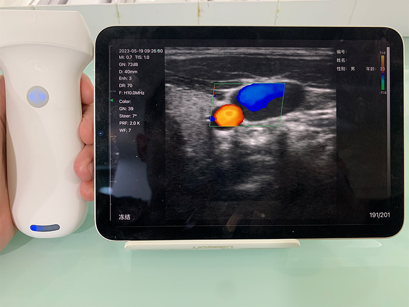
Vascular access and dialysis. This is where the probe has the strongest field record. Haidian Hospital's nephrology department was reported as an early adopter of wireless pocket ultrasound for arteriovenous fistula cannulation, and the vascular surgery team at Peking University First Hospital performed what was described as China's first wireless portable ultrasound-guided endovascular treatment of a dialysis access. Both rely on the same feature: real-time guidance in a sterile field with no cable in the way.
Emergency and critical care. Xianyang First People's Hospital used a wireless probe to guide pericardial effusion drainage in a 79-year-old high-risk patient — a bedside procedure where moving a cart into position is part of the problem.
Teaching, line services and telemedicine. Jiangsu Province Hospital documented wireless ultrasound in PICC placement, and Xiangxi Tumor Hospital used it for catheter placement in difficult-access patients. Streaming to several screens at once turns a single scan into a teaching moment or a remote consult without extra hardware.
Veterinary and non-clinical use. Animals do not hold still for a trolley. The company also states an intention to position the convex 3D probe as a “fetal camera” for non-medical settings such as family use, maternal retail and studios — a roadmap claim worth quoting carefully rather than treating as an approved indication.
FAQ
Is the volume real or interpolated from 2D? It is reconstructed from a spatial sweep and stored as voxels. Orthogonal sectioning, in-volume measurement and DICOM export are the practical proof.
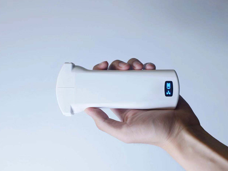
Does a heavier probe mean worse ergonomics? At 325 g it is heavier than a 200 g 2D unit because of the sweep mechanism, and still light enough for single-hand use against a patient.
What training does a 3D workflow need? A sweep takes seconds and reconstruction is automatic. The skill sits in choosing the plane that answers the clinical question — the same skill a conventional 3D system demands.
Guangzhou Sonostar Information Technology Co., Ltd. is a high-tech enterprise specializing in miniaturized, intelligent ultrasound. We handle R&D, manufacturing, sales and service in-house, with a product line built on probe-based handheld ultrasound imaging systems and wearable scanners. Our devices are CE- and FDA-registered, exported to over 100 countries, and used across clinical care, primary-care emergency response, aesthetic medicine and veterinary practice — bringing ultrasound within reach of more patients. Happy to discuss any requirement or question you may have.

 Home
 Product
 News
 Contact